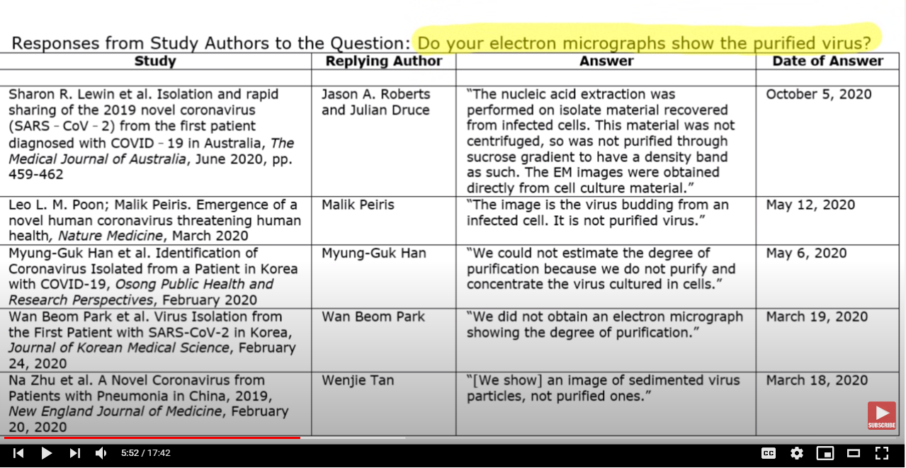
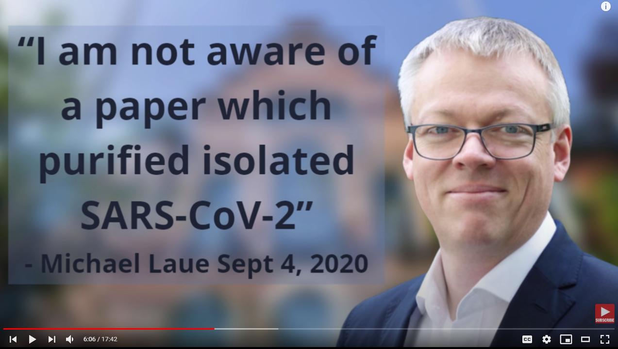
The whole you-didn't-image-this-in-a-highly-purified-sample is a red herring with regard to determining whether a (suspected) virus is a "a brand new viral pathogen" or not. Nobody does that kind of novelty determination based on imaging the virus... simply because lots of viruses look alike even under electron microscopy. Imaging is not a useful way to tell apart viruses from similar "genres", e.g. various coronaviruses one from another. I didn't expect there to be a paper about this, but (amusingly) there is (a short) one that's somewhat relevant, starting with:
Caution in Identifying Coronaviruses by Electron Microscopy
We are concerned about the erroneous identification of coronavirus directly in tissues by authors using electron microscopy. Several recent articles have been published that purport to have identified severe acute respiratory syndrome coronavirus 2 (SARS-CoV-2) directly in tissue. Most describe particles that resemble, but do not have the appearance of, coronaviruses. [...]
(I'm not going to delve into who is right in that little spat, just saying that identification by imaging is much more problematic, and has been superseded by molecular methods by and large.)
Furthermore, while the video lambast at length PCR tests as a diagnostic tool, it's completely silent on how suspected new viruses are actually sequenced in the first place. There is a general rant against molecular biology in the final minutes of the video, but this is still unspecific. That rant is basically akin to: we can never make/understand computers with billions of transistors because of their complexity.
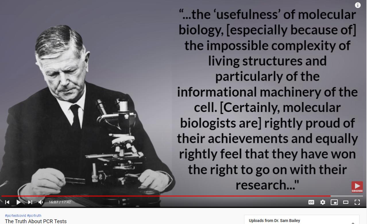
N.B.: that's a quote from a 1966 position paper of Macfarlane Burnet. The same author in a 1970s book "predicted that scientific progress would end soon", according to Wikipedia. His beef/prognostication generally was with DNA/RNA-based identification (which wasn't actually possible during his active career).
Basically, Dr. Bailey doesn't (explicitly) outline a single method by which she would agree that something is a new pathogen. So to a large extent, the whole video is an argument that we can never know this.
And while Burnett may have railed against molecular methods, the micrographs he used in his own papers on influenza don't seem to ever show the purified influenza virus alone, but commonly show it among cells. E.g., these are images from Burnett's 1957 article in Sci. Am.
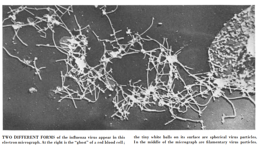
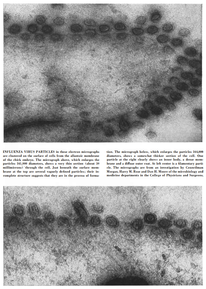
So really, this purification before micrographs is a Bailey-made-up standard that Burnett himself probably would have not considered seriously.
Now to save this answer from triviality, the way by which suspected new viruses are sequenced is a bit different than what (later) PCR diagnostic tests do... because the latter use highly specific primers, which aren't known (what they should be) when sequencing an unknown virus in the first place. An umbrella term for such methods is metagenomic sequencing.
If you look at actual (NEJM) paper for SARS-CoV-2 discovery, they mention using Illumnia and nanopore sequencing specifically. They don't bother citing any publication for these methods, presumably because they are considered (already) well known, although nanopore sequencing is actually kinda new.
The NEJM paper on SARS-CoV-2 identification doesn't detail much purification steps (it's a brief report) but does mention that they excluded known respiratory pathogens with standard kit for these (details are in a supplement to the paper). Simplifying, the logic of the paper is: these patients have these symptoms of a respiratory disease, they test negative for (a whole lot of) known pathogens that can cause these symptoms, and looky we found a novel virus sequence in their throats; ergo, that must be the cause of their disease. I'm simplifying, because they also cultured the new pathogen etc. and used control samples of the culture without the pathogen:
Extraction of nucleic acids from clinical samples (including uninfected cultures that served as negative controls) was performed with a High Pure Viral Nucleic Acid Kit, as described by the manufacturer (Roche)
If you read the mfg description
The High Pure Viral Nucleic Acid Kit is intended for general laboratory use and efficiently purifies viral nucleic acids from serum, plasma, or whole blood using sample volumes of up to 200 μL. The purified viral nucleic acid is eluted in nuclease-free water, and is suitable for direct use in PCR and RT-PCR.
An earlier (2015) paper on nanopore sequencing for example (which compares it to Illumnia) also does mention some purification steps
cDNA were purified using AMPure XP beads [...]
An even earlier 2013 review on viral identification goes into some nitty gritty details on some purification steps, e.g.
Pathogen enrichment or host depletion before microarray and deep sequencing analyses hence becomes critical to maximize sensitivity for identification of novel agents in clinical samples. For viruses, capsid purification procedures involving repeated freeze/thaw cycles, filtration, ultracentrifugation, and prenuclease digestion have been developed to enrich host tissues or body fluids for infectious particles. Strategies to deplete the sample of background host DNA can also be implemented, including the use of methylation-specific DNAse to selectively degrade host genomes, removal of host ribosomal RNA, and/or removal of the most abundant host sequences by duplex-specific nuclease (DSN) normalization. Another complementary approach is to perform target enrichment using biotinylated probes to enrich NGS libraries for sequences corresponding to pathogens, akin to now well-established techniques that have been developed in the cancer field. [...]
Which steps are relevant (for which method) may a good question on Biology SE, but Dr. Bailey seems completely ignorant of any of this, even on a high level, at least in the video. Basically, Dr. Bailey's rant is that you can't purify some RNA or resultant cDNA (which isn't present in control cell cultures), you need to "purify the [whole] virus". This is nonsense from the point of view of practitioners in field of new virus discovery.
As far as Covid-19 is concerned largely similar approach is taken in Shi et al.'s Nature paper: metagenomic sequencing, followed by a general confirmation with electron microscopy.
I'm gonna close this by saying that sequencing is still a fast moving field, technologically speaking. Methods that were used e.g. in 2003 to identify the SARS virus are only relevant nowadays as a general idea... but even that paper mentions:
nucleic acids were purified from the supernatant. Random amplification was performed with 15 different PCRs under low-stringency conditions. We had previously shown that this method is able to detect unknown pathogens growing in cell culture (unpublished data). To detect RNA viruses, an initial reverse-transcription step was included.
Basically someone from outside the field who rants that the field is invalid to begin with because of its complexity and also complains that some (almost certainly not needed) whole-virus purification steps aren't performed to their idea-of-a-standard is basically (almost) as laughable as the average guy on the street complaining that some mathematician isn't writing proofs according to Trump's speeches.
What gives a high degree of confidence in the sequencing results regarding SARS-CoV-2 is also the fact that the sequencing process (and I'm not talking about PCR testing with specific kits but basically a replication of the initial analysis/experiments be it with various sequencing technologies) for has been done for more than 90,000 samples, worldwide. Basically that's an experiment/analysis replicated 90,000+ times. This wasn't done because the initial results were deemed not credible, but mainly because viruses mutate/evolve and establishing lineages and keeping track of mutations which may affect is important for epidemiologist too. (See e.g. an early mutation that enhanced transmissibility being identified; or lineages of concern because e.g. they have some antigenic mutations, meaning they "work around" some antibodies hosts may have against other lineages.)
Purified whole-virus samples are actually used in some studies. For example there's one such study on mouse hepatitis virus (which also a coronavirus), but the goal there was to measure the variation in particle size and likewise for their wall thickness and spike lengths. For something like that, it's convenient to have a bunch viral particles in close proximity so you can easily measure a set of them from a few images.
Perhaps with all that methods stuff, I didn't make some basic science clear enough. You can recreate a virus' capsule (thus the "whole virus") solely from its nucleic acid, by injecting the latter into an appropriate host cell. The virus will replicate inside the cell and eject from the cell new, encapsulated viral particles. (More technically, that viral capsule is called a capsid.) Some viruses--including coronaviruses-- are additionally coted with a lipid layer, which is also obtained in roughly the same way, although a bit later in the cell-egress process. (Viruses which cause the cell the to burst [aka lysis] don't get this additional envelope.)
Such "capsule recreation" was done e.g. with Covid-19's virus (SARS-CoV-2). Not only that, but the whole nucleic acid of the virus was created "from scratch" in that (last) experiment, simply following the "recipe" given by its published sequence. (This kind of tech has existed for about two decades.)
This is why the capsule is irrelevant to the identity of the virus.
Not only that, but as an aside, the same virus can have different envelopes, e.g.
Hepatitis A virus (HAV) spreads by both the fecal-oral route (by ingestion of food contaminated with fecal matter) and the blood route (such as by use of contaminated needles). HAV can have both naked and quasi-enveloped capsids. We know that HAV particles isolated from fecal matter are naked, while particles circulating through the blood are protected by a quasi-envelope that prevents detection of these particles by the host immune system.

